$3.8M NIH grant funds 3D tissue and organism visualization project
Keith Cheng, distinguished professor of pathology, of pharmacology and of biochemistry and molecular biology at the Penn State College of Medicine, has been awarded a four-year, $3.8 million grant from the National Institutes of Health to further develop novel methods for 3D imaging tissue.
Cheng, who is also affiliated with the Huck Institutes of the Life Sciences, Penn State Cancer Institute and Institute for Computational and Data Sciences, said he plans to broaden the new form of 3D imaging his team has developed, which he called “X-ray histotomography,” to characterize all tissues and cell types. Cheng’s efforts will focus largely on whole traditional models of human biology and disease, such as fruit flies and zebrafish. His team will also image parts of larger organisms like mice and humans.
Like computed tomography (CT) scans, Cheng’s technique uses X-ray beams to take sets of pictures at thousands of angles in two dimensions (2D). These sets of 2D images are then converted into 3D images to create models of plant and animal samples up to a centimeter in diameter.
“We are creating models of the entire nervous system in a whole animal from a single 3D image captured in about an hour — a first,” Cheng said.
This new technology promises potential advancements in cancer diagnosis, according to Cheng, as diagnoses rely on the microscopic study of all the cells and their arrangements in pathology tissue samples. In fact, his lab’s project, “Building a Wide-field, High-resolution Histotomography Resource for Biology,” was inspired by a problem in cancer diagnosis.
“For practical reasons, we study only a small percentage of the original sample; furthermore, we are limited to slices that typically represent only a fraction of each cancer cell,” Cheng said. “This is enough to recognize tumor type, but we cannot measure their cell shape or volume, or evaluate all the cancer cells and their variants in a tumor. In the early 2000s, I realized that new lenses, imaging chips and computer power would make possible the dream of 3D imaging whole cancer samples at histological resolutions. … I asked, ‘How do we use new technology to create 3D images of centimeter-sized tumor samples and organisms, to allow pathologists to diagnose more precisely?’ More broadly, ‘How do we automate the detection and measurement of every cell and its arrangements in any tissue in any organism?’”
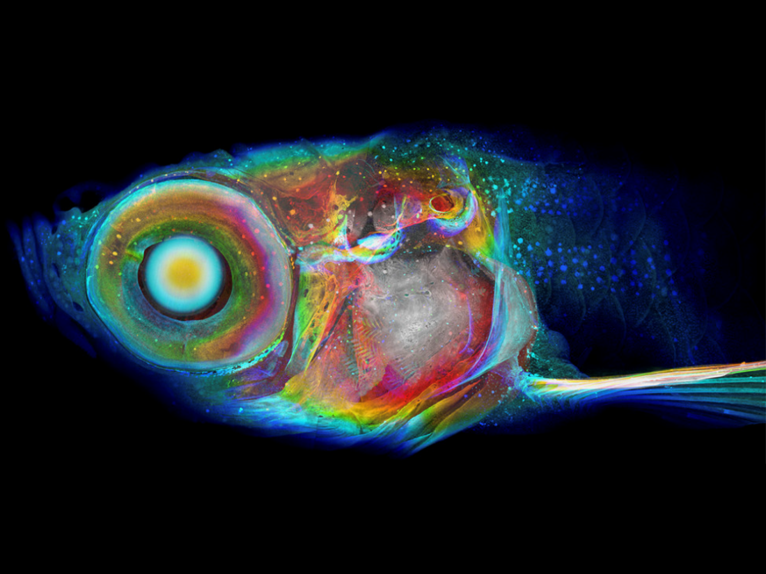
Knowing that pathology samples are the same size as many model organisms, Cheng’s interdisciplinary team developed a way to image entire small organisms, starting with zebrafish and tissue samples. The 3D computational images would allow virtual slices at any desired thickness of whole, intact samples at any angle, ultimately allowing scientists and physicians to measure cell shape and size and their arrangements in tissues and organisms.
Initial attempts to use series of physical slices to create 3D images proved impractical, leading Cheng to rethink the project and experiment with the use of X-rays to accomplish the goals.
“In 2019, we published the first paper showing that you could see the 3D structure of every tissue in a whole vertebrate organism, the zebrafish, using customized X-ray imaging that utilizes the same principles as human CT, but at 2,000 times the resolution,” Cheng said.
This enabled Cheng and his colleagues to reach the previously impossible goal of achieving high enough resolution to recognize cell types and cellular change in any part of entire centimeter-scale specimens. In a study published in 2022, the team demonstrated micron resolution at a half-centimeter scale. Now, the team has achieved similar resolution at centimeter scale.
“Developing imaging of centimeter wide volumes at voxel, or 3D pixel, resolution of less than a micron may not sound like much, but that’s a major advance in the 3D imaging world,” Cheng said. “You can normally image a large sample at poor resolution, or a small sample at high resolution. Now we can accomplish both. We are now working to democratize the imaging and digital dissection of 3D models, so that we can pioneer ways to analyze the cells and their relationships — goals of the Penn State computational phenomics initiative.”
“Dr. Cheng’s vision and leadership in the field of advanced imaging is an asset not only to our College of Medicine, but science as a whole,” said Karen Kim, dean of Penn State College of Medicine. “By providing unprecedented insights into the intricate details of tissues and cells, this research has the potential to revolutionize the field of medical imaging and significantly impact the diagnosis and treatment of cancer and other diseases.”
The research team employs electron accelerators known as synchrotrons at the Argonne National Laboratory (ANL) near Illinois and the Lawrence Berkeley National Lab (LBNL) in California. These powerful X-ray sources, with Cheng’s unique imaging system, create high resolution CT-scans at larger scales than were previously possible.
“The intensity and ‘purity’ of synchrotron X-rays mean that they provide the fastest way to get 3D images of large tissue samples at high resolution,” Cheng said. “Using the synchrotron is equivalent to using the sun versus a night light for photography.”
Retaining resolution for larger samples imaged required the researchers to advance camera lens design and rendering software. Physicist Steve Wang at Mobile Imaging Innovations, Inc. developed the Cheng lab’s lens systems; Patrick La Riviere, professor of radiology at University of Chicago and co-investigator on the grant, is developing helical reconstruction approaches to create single CT images without needing to stitch together the results of separate thinner scans. Sharon Huang, professor of information sciences and technology at Penn State, is helping the team to apply the growing power of artificial intelligence (AI) to increase image quality and accelerate analysis possible.
Additional collaborators include physicists at ANL and LBNL and biologist and geneticist Khai Chung Ang, assistant professor of pathology and scientific director of the zebrafish research facility at Penn State.
“In the life sciences, the development of new technologies to visualize the structure and function have always been a catalyst for new discoveries and new ways of thinking,” said Patrick Drew, interim director of the Huck Institutes of the Life Sciences. “We are very excited by the promise of this technology and are looking forward to the new vistas it opens up.”
The team is currently crafting ways to democratize visualization and analysis of these 3D scans for the scientific community and said they are planning further collaborations with other experts to develop applications intended to accelerate work across a wide swath of disciplines.
“This is a dream interdisciplinary project with applications across biology, medicine, agriculture and AI and involves collaboration with industry and multiple government agencies,” Cheng said. “I love to keep asking, ‘What of great importance can we accomplish together that none of us can do alone?’”
For a deeper dive into this technology, listen to this November 2022 episode of the Researchpod podcast, “X-ray histotomography: Characterizing every cell of a whole organism.”
If you're having trouble accessing this content, or would like it in another format, please email Penn State Health Marketing & Communications.