Calcium in breast arteries predicts future cardiovascular disease
New study findings suggest that routine mammograms could be a tool for evaluating cardiovascular disease risk in women
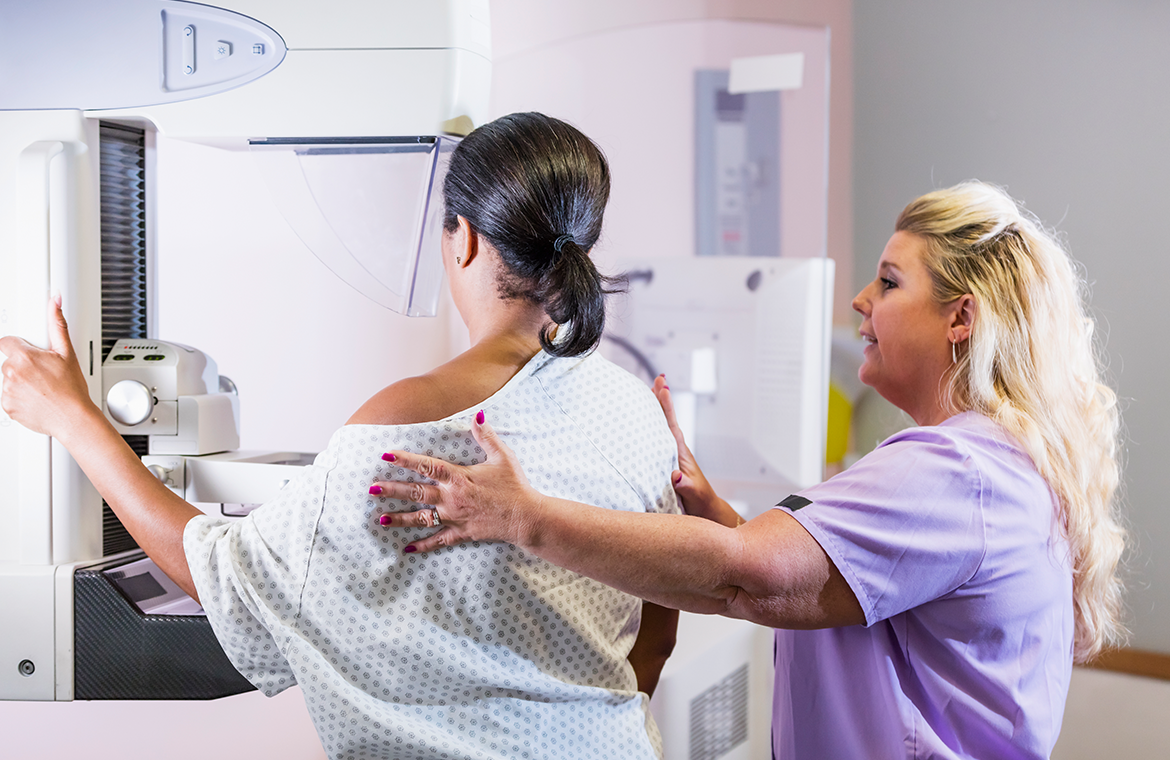
Routine mammograms are a critical tool for breast cancer screening. However, they may also hold crucial, potentially untapped information about a person’s risk for cardiovascular disease, the number one cause of death among adults. The X-ray images can detect calcium in the breast’s arteries, a sign that the blood vessels are getting stiffer.
New research — presented on Dec. 3 at the Radiological Society of North America meeting by Matthew Nudy, assistant professor of medicine and public health sciences at Penn State College of Medicine — found that the severity of calcium accumulation in breast arteries and the progression of this calcification seen on mammograms predicted future cardiovascular disease. In this study, researchers found that women with more severe calcification and with calcification that progressed over time had an increased risk for major events, like heart attack, stroke, heart failure and death.
These findings suggest that breast arterial calcification could be a marker for cardiovascular disease and may help identify women who are at greatest risk for cardiovascular disease.
“We know that women are more likely to be diagnosed at later stages of cardiovascular disease and have worse outcomes following a heart attack compared to men. That may be in part because the current cardiovascular risk assessment tools underestimate risk in women. We need better tools,” Nudy said. “In the future, assessment of breast arterial calcification may help improve our ability to predict risk and prevent cardiovascular disease.”
As people get older, calcium can collect in the arteries, which increases the risk of heart attack and stroke, explained Nudy. For some patients, physicians may recommend a CT scan to determine if there is calcium build up in the coronary arteries, the blood vessels that supply the heart. There are downsides to the test, Nudy noted, that might keep patients from pursuing the test and physicians from recommending it, including costs and exposure to radiation from the scan. Conversely, mammograms, which can pick up calcifications in the walls of arteries of the breast, are already widely used for breast cancer screening, with both the American Cancer Society and the United States Preventative Services Task Force recommending at least biennial screening in women starting at age 40.
Currently, the presence of calcium build up in breast arteries isn’t routinely included in radiology reports since there’s no known association with breast cancer. However, previous studies, including a systematic review and meta-analysis led by Nudy, have found that breast arterial calcification is associated with future cardiovascular disease and death.
In this study, the research team analyzed data from 10,348 women from an academic medical center in the United States who had sequential mammograms, with an average of 4.1 years between mammograms. The average age of participants was 56. For each mammogram, the team used an investigational version of the cmAngio artificial intelligence (AI) software — developed by CureMetrix, a company that tests and advances AI tools for medical imaging analysis and partnered on this study — to determine if calcification was present in breast arteries and its severity. The model generated an adjusted score based on the length of the artery calcification. Participants were then separated into four age-adjusted categories of breast arterial calcification severity based on their score: negative, mild, moderate and severe.
The researchers found that vascular calcification was present in 19.4% of participants at baseline. Those with more calcium accumulation in the arteries over time had increased risk of a serious heart-related event — up to two times higher risk for those in the severe score category.
The study, one of the first to look at breast arterial calcification progression over time, also showed that calcification can worsen over time, occurring in as little as a year. The faster the profession occurred, the greater the cardiovascular risk, too.
Those who didn’t have calcium in their breast arteries on the initial mammogram showed the lowest risk of progression, but if calcium was detected on a follow-up mammogram, they had a 41% higher risk for an adverse cardiovascular event and death over an average of 5.6 years of follow-up. For those who started in the mild category and progressed to any higher category, they had a 59% higher risk. For those who started in the moderate category and progressed to the severe category, they had a 93% higher risk.
“This could be a way to use data that may already be available for different reason and to potentially use it to risk stratify an individual for the development of cardiovascular disease,” Nudy said. While the findings suggest that calcification of the arteries in the breast is a warning sign for cardiovascular disease, he cautioned, more research is needed to understand this relationship and how the information could be used by clinicians.
Other collaborators on the research include Nitesh Nerlekar, associate professor and deputy director of the Victorian Heart Institute, Monash University and Alyssa Watanabe, adjunct clinical associate professor at the University of Southern California. Richard Mantey, Junhao Wang and Homa Karimabadi from CureMetrix also contributed to this work.
For studies presented at the Radiological Society of North America meeting, there is a double-blind peer-review process.
This work was supported by CureMetrix.
If you're having trouble accessing this content, or would like it in another format, please email Penn State Health Marketing & Communications.