Medical student uses 3D printer to create ultrasound “phantoms”
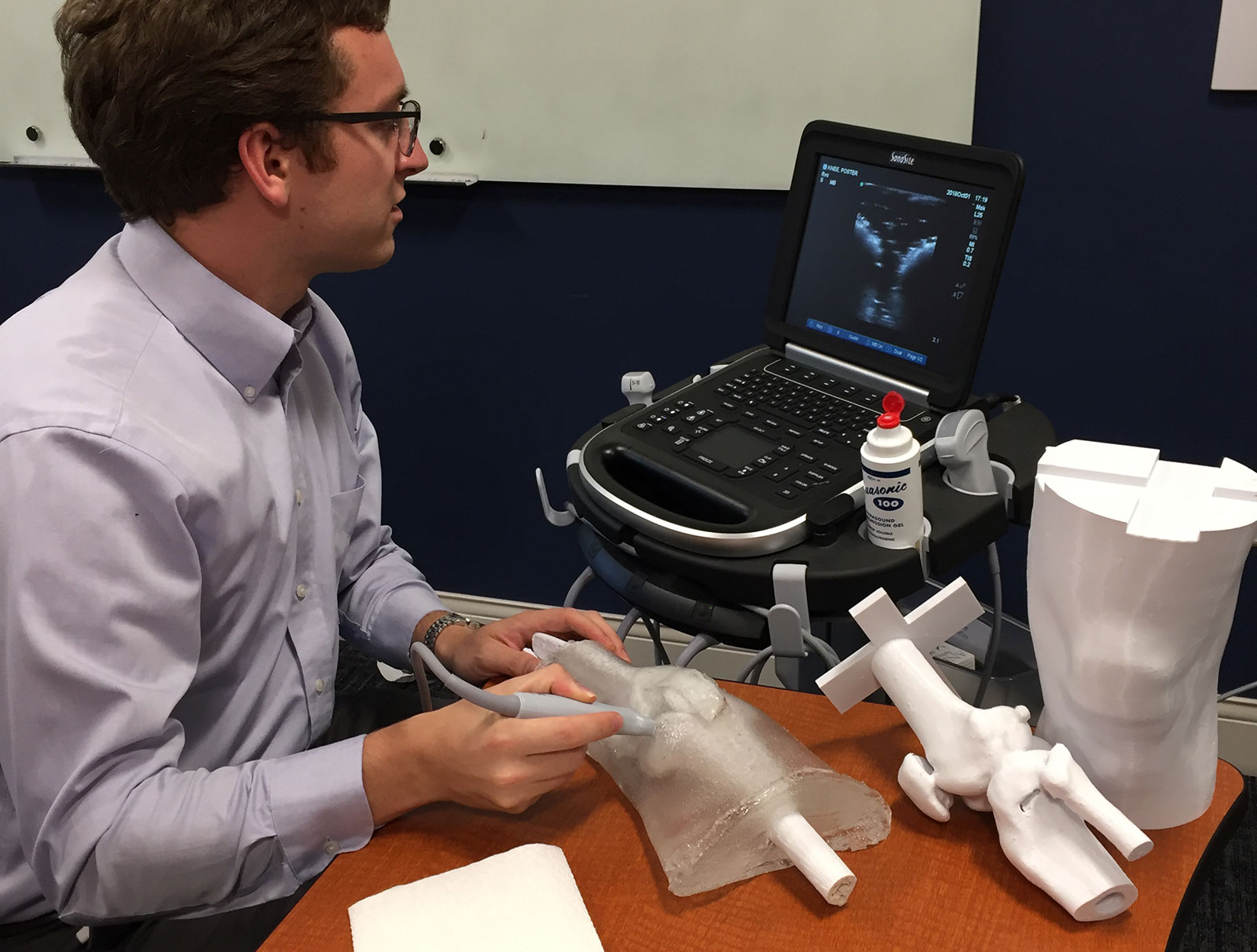
The 3D printers in the Technology Innovation Sandbox at Harrell Health Sciences Library have been involved in several interesting projects during the past couple of years, and medical student Jason Spicher’s project is a good example. Spicher has not only used the 3D printers, but has also used the library’s copy of Mimics to help create some of the models to be printed. Using hard plastic for bone and ballistic gel, Spicher has started working on a project that could help to train future students to perform ultrasound-guided joint injections.
“In order to train providers to perform ultrasound-guided joint injections, you need a phantom, a model that looks like human anatomy when viewed with ultrasound,” Spicher said. He went on to note that, though commercial ultrasound phantoms are available, they can be quite costly. “I am trying to make a model that can be made inexpensively and used by academic medical centers around the country,” he said.
3D printing technology has been at the core of his project. Spicher said he has been able to make quick and inexpensive mock-ups of the skeletal component, in addition to quickly adjusting the skeletal pieces to fit within the purposes of his phantom. He has used Mimics to turn CT and MRI scans to 3D models, ready for printing, as well as software such as TinkerCAD to make minor adjustments to the models.
Spicher has had to work through a couple of obstacles. “One issue I’m working with now is that the 3D printed parts are hollow,” he said. “This saves material and money, but when they are placed in the leg mold with the molten ballistic gel, they rise to the top because they’re less dense. This caused the whole production process to speed up.”
Since the ballistic gel didn’t cool correctly, it caused bubbles on the surface of the 3D printed pieces. This can interfere with the function of the phantom, so Spicher used modeling software to design a new support structure that will keep the bones stationary, allowing the gel more time to cure, removing all of the bubbles.
Spicher’s goal is to streamline the production of the project so that someone from another medical center could build it within a day using his plans and 3D files. After that, he plans to move on to other joints, such as the ankle.
Read more
If you're having trouble accessing this content, or would like it in another format, please email Penn State Health Marketing & Communications.