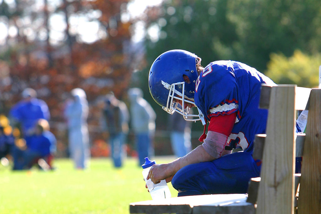
Summer 2019 edition: Sports Medicine for the Primary Care Provider

The Summer 2019 edition of Sports Medicine for the Primary Care provider was recently published. Articles in this edition include “The Performing Arts Athlete and Overuse Injuries,” “Chondrosis Treatment Varies by Cartilage Defect, Patient Symptoms” and “Research in Brief.”
A printable version of this newsletter is available here.
All articles also appear below.
Welcome
Dear Fellow Health Care Provider,
I hope you are enjoying the great outdoors this summer and having a safe and injury-free time! Welcome to the latest edition of our primary care sports medicine newsletter, a biannual publication focusing on seasonal sports topics. We hope you find the information useful and appreciate any feedback you have to enhance our efforts.
For questions, feedback and inquiries about future issues, please contact Tracie Kirkessner at tkirkessner@pennstatehealth.psu.edu or me at msilvis@pennstatehealth.psu.edu.
Enjoy,
Matthew Silvis, MD
Professor, Departments of Family and Community Medicine and Orthopaedics and Rehabilitation
Penn State Health Milton S. Hershey Medical Center

The Performing Arts Athlete and Overuse Injuries
By Kileigh Hess, MS, LAT, ATC, PES
The performing arts athlete, just as any other athlete, needs strength, power, flexibility, functional movement and core stability to be at their peak performance. However, dancers are more likely to suffer injuries compared to a traditional sports athlete. Research has shown that 80% of dancers have at least one injury per year that affects their ability to perform – compared to a 20% injury rate for football players. Since dance is not a contact or combative sport, and these performers are not participating in a competitive competition on the stage, why are they suffering such a higher rate of injury?
Overuse injuries are the most common injury in the performing arts setting with a majority of injuries affecting the lower extremity. Compared to acute injuries, overuse injuries are more subtle and occur over time. They are the result of repetitive microtrauma to tendons, bones and joints. Examples of injuries that occur in dancers include, hip flexor tendonitis, hip bursitis, SI joint dysfunction, Achilles tendonitis, patellar tendonitis and patellofemoral pain syndrome. Overuse injuries in dancers typically present as pain.
There are many more factors that predispose performing arts athletes to overuse injuries. One major factor is that there is never a true “off-season.” On average, performing arts athletes spend five hours a day practicing at high rates and speed, which leaves them little time to recover between sessions. More external factors include poor technique, performance space, costumes, props and shoe type and fit. While some of these factors can be changed, personal factors that can impact injury also include foot type, body alignment, joint stability and strength. Restrictive diets and unhealthy body weights may also contribute to these injuries. Proper nutrition is important for dancers of all ages. There are sex-specific differences as well between male and female dancers. Males are usually expected to meet tougher athletic requirements, whereas females tend to have more demanding technical requirements, meaning that females are affected more by overuse injuries.
Proper posture is important along with cross-training and physical therapy to help aid in the prevention of overuse injuries. Cross-training helps to build strength and endurance throughout the body. Core and hip strengthening exercises that are stability- and strength-based are ideal, along with aerobic and cardiovascular activities. However, rest is the most important way to prevent overuse injuries. It is recommended that at least two days off in a row are taken after the most intense training in the weekly schedule. Once a season or show is finished, a three- to four-week period of rest is best for recovery.
With proper education, treatment, guidance and an established relationship with the performing arts athlete, the health care professional can help abolish the myth that performance-related discomfort is inevitable and that the problem will eventually fix itself.
Chondrosis Treatment Varies by Cartilage Defect, Patient Symptoms
By Robert Gallo, MD
“Chondrosis” involving a joint surface is one of the most common findings discovered on magnetic resonance imaging of a joint. In an era where patients have access to imaging reports, chondrosis can become a major area of concern and confusion for patients and providers alike. The purpose of this article is to discuss some of the important concepts of chondrosis and provide some strategies to guide the primary-care physician.
“Chondrosis” literally means “cartilage genesis” or growth, but is commonly considered to be degeneration of the cartilaginous surface lining the ends of an articulating bone. In healthy joints, this cartilaginous surface is composed of hyaline cartilage, a surface that is smoother and produces less friction than ice. When subjected to injury or deterioration, hyaline cartilage does not have the capacity to heal or repair itself once the growth plates are closed during adolescence. Instead, the hyaline cartilage wears away or is replaced with fibrocartilage, an inferior and less durable form of cartilage.
Chondrosis can be found in varying degrees which are categorized by the depth of the cartilage lesion. The most accepted classification to define the degree of chondrosis in a given region was described by Outerbridge. This classification system was originally created to describe chondrosis arthroscopically but has been adopted for use in describing MRIs (Table 1). While this system does help stratify level of chondral injury or degeneration, there is considerable difficulty differentiating Grade 2 from Grade 3 lesions. Treatment of chondrosis varies widely depending upon location and size of lesion, depth of cartilage defect and patient symptoms. Nonoperative measures such as physical therapy, medications, bracing and injections are mainstays of treat for Grade 1, 2 and most Grade 3 lesions. While corticosteroid injections can be successful in relieving pain associated with chondrosis, many physicians believe that corticosteroid injection may hasten the cartilage deterioration, especially with repeated injections. Therefore, platelet-rich plasma and viscosupplementation, both agents thought to be less deleterious to the articular cartilage, are popular injection options for treating low-grade lesions.
To date, orthopaedists have struggled in attempts to recreate articular cartilage. Most surgical techniques are reserved for focal Grade 3 or 4 chondral lesions affecting only one location within a joint. “Kissing lesions,” or those lesions involving both sides of a joint, are generally considered to be nonamenable to cartilage restoration. Current popular options for cartilage restoration include microfracture, osteochondral autograft or allograft, and autologous chondrocyte implantation (ACI). A description for each is contained in Table 2. Microfracture remains because this technique yields only a hyaline cartilage fill, while osteochondral autograft lesions are limited by size of the lesion, i.e., there is only so much cartilage that can be transferred without causing donor-site morbidity. Osteochondral allograft and ACL are limited by costs. Total knee replacement, which replaces the articular surface with plastic and metal, is reserved for those with extensive Grade 4 chondrosis.
Table 1: Arthroscopic and MRI Findings in Various Stages of Chondrosis
Table 2: Cartilage Restoration Options

Research in Brief
Discrepancies in Child and Parent Reporting of Concussion Symptoms
Authors: Ruikang Liu, MD, and Steven Hicks, MD, PhD
Affiliation: Penn State College of Medicine, Department of Pediatrics, Hershey, Pa.
Purpose: The Sports Concussion Assessment Tool (SCAT) is one of the best-established multidimensional tools for sideline assessment of sports-related concussions. A component of this tool is the Post-Concussive Symptom Inventory (PCSI), which is a symptom checklist that can be filled out by the child and parent to characterize subjective symptoms following a concussion. However, the consistency between child and parent responses is unclear, so the purpose of this study is to assess the differences in child and parent reporting of symptoms following a concussion.
Methods and study design: Prospective cohort study. Enrolled patients age 7-21 diagnosed with a concussion within the last two weeks. Child and parent filled out the PCSI of the Child SCAT-3 at the initial evaluation (61 respondents) and four weeks post-injury (30 respondents). A two-tailed T-test was used to compare response differences in 1) individual symptoms (scale 0-3); 2) total number of symptoms (out of 20); and 3) total symptom severity burden (out of 60 possible points).
Results: At the initial visit, there was a significant (p < 0.05) difference between child and parent report of symptom severity for "distracted easily" (p=0.001), "confused" (p=0.001) and "forget things" (p=0.035). Parents underestimated the severity in each of these relative to child-report. At four weeks post-injury, there was a significant difference between child and parent report of symptom severity for "problem remembering" (p=0.026) and "forget things" (p=0.045). At this timepoint, parents overestimated the severity of these symptoms relative to child report. The total number of symptoms reported by children at four weeks post-injury (4) was also lower than the total reported by parents (5, p=0.047). Conclusions: Cognitive symptoms (e.g., distraction, confusion, memory) most frequently showed a difference in reported symptom severity when comparing child and parent reports at the initial concussion evaluation. This trend continued four weeks post-injury. Parents underestimated symptom severity at the initial visit and overestimated severity and total number of symptoms four weeks post-injury.
Significance: Severity of cognitive symptoms in concussion may be perceived differently by children and parents. This finding underscores the need for objective assessments of cognition in children with concussion, which may aid return-to-learn management.
Acknowledgements: Special thanks to the Children’s Miracle Network for its funding. Additional thanks to Andrea C. Loeffert, DO; Jeremiah J. Johnson, MA, BS; Jennifer Stokes, BS; Robert P. Olympia, MD; Harry Bramley, DO; Molly C. Carney, BS; and Neal J. Thomas, MD, MSc, for their contributions in early project design, writing and data collection.

How Effective is the Musculoskeletal Screen During Mass Pre-participation Physicals?
Authors: Stephanie Carey, MD, MPH; Sandy Zettlemoyer, DAT, ATC; Matthew Silvis, MD; Ming Wang, PhD; and Cayce Onks, DO, MS, ATC
Affiliations: Penn State Health Milton S. Hershey Medical Center (Carey, Silvis, Wang and Onks); Mechanicsburg School District (Zettlemoyer)
Purpose: There is concern that the current format of the Pre-participation Physical Examination (PPE) is not efficacious for identifying individuals who may be at risk for athletic-related injury or illness. The purpose of this study is to review the effectiveness of the musculoskeletal (MSK) screening examination from the 4th ed. PPE monograph by comparing disqualification and injury rates between standardized and control groups.
Methods and study design: Retrospective review of two PPE sessions for middle and high school students during the 2017-2018 school year. Athletes attending the first offered mass PPE had an MSK screening exam standardized across all providers. Group 2, the control, were athletes who attended the second mass PPE where providers completed the MSK exam of their choice. The rates of disqualification were analyzed. Injury rates were then calculated from the entire year following their PPE.
Results: 152 standardized and 234 nonstandardized MSK exams were performed. There was no significant difference in the rate of disqualification between the standardized and control groups respectively (0.40 (4/10) and 0.60 (6/10), p=0.53). There was no significant difference in the injury rates (calculated as injury incidence proportion) between the standardized and nonstandardized group respectively (0.263 and 0.192, p=0.15).
Conclusions: The current recommendation to include a standardized MSK exam is not superior to a provider’s choice MSK exam for detection of disqualifying injuries or conditions that predict injury.
Significance: Current MSK screening recommendations should be reviewed for their ability to effectively meet their intended goals. Future research should evaluate how we teach the MSK portion of the PPE and if there are opportunities to improve the content of the MSK screen.

Contact Us
Learn more about the Sports Medicine team at Penn State Health here.
If you're having trouble accessing this content, or would like it in another format, please email Penn State Health Marketing & Communications.
