Cheng receives NIH funding for next phase of 3D imaging project
A Penn State-led research project to develop a new, diagnostic, three-dimensional imaging technique will continue thanks to a four-year, $2.6 million grant from the National Institutes of Health (NIH). If the researchers succeed, the method could replace 2D histology, the current state-of-the-art method of studying cells, tissues and organs.
The researchers say this could impact drug development, diagnostics and basic understanding of how genes and environment define phenotype, an organism’s observable characteristics and traits.
Dr. Keith Cheng, distinguished professor of pathology and laboratory medicine, biochemistry and molecular biology and pharmacology at Penn State College of Medicine, will work with Sharon Huang, associate professor of information sciences and technology at University Park, on the project. They explained that histology requires pieces of tissue to be sliced into thin, two-dimensional slices – a technique that only uses less than 2 percent of a specimen and yields no three-dimensional data.
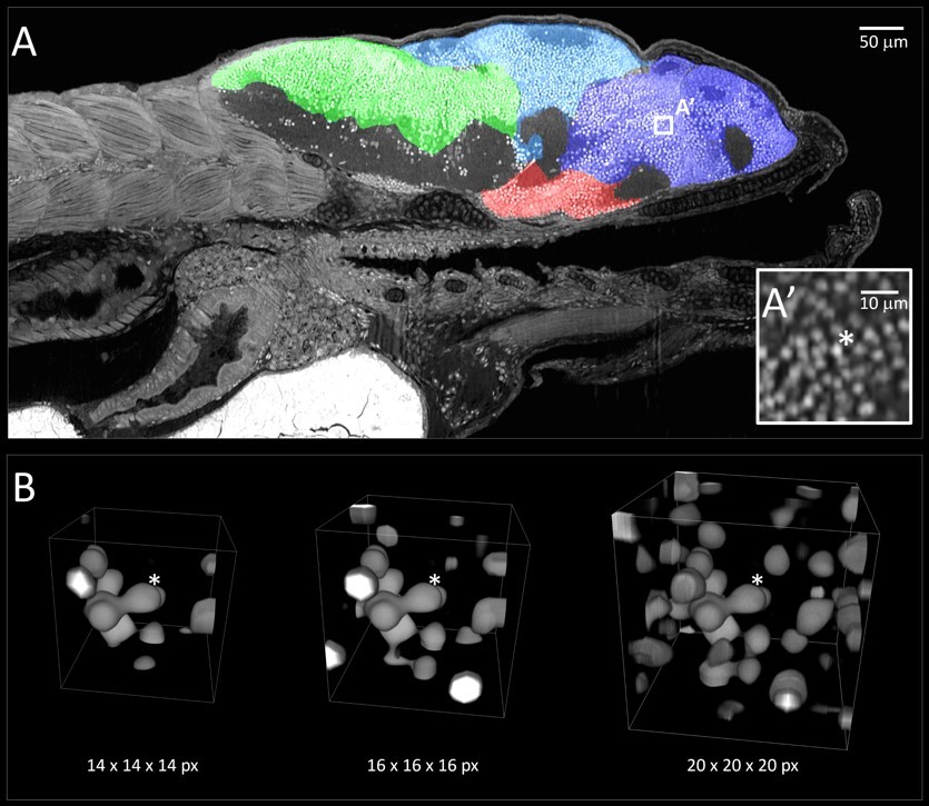
Cheng’s lab showed in previous work that X-ray “histotomography,” built on the basic principles used in CT scans, can be used to create 3D images from mature and pigmented samples at resolutions one thousand times greater than CT scans. The volume data can be used to visualize structures in 3D.
The funding received from the NIH grant will allow the researchers to apply principles of biology, physics and machine learning to improve image resolution, processing speed and analytics.
Read more about the next phase of this project on Penn State News.
If you're having trouble accessing this content, or would like it in another format, please email Penn State Health Marketing & Communications.